Monday, 28 November 2011
Monday, 21 November 2011
Tuesday, 15 November 2011
MANAGEMENT OF SOME TEETH TRAUMA
By Augustine Mbehoma Rukoma
1. Intrusive luxation with ankylosis-
· orthodontic uplifting may be done
2. Treatment of tooth fracture at or below cervical level
· Retain coronal part using luting cement and post
· Extrude the tooth orthodontically in order to bring the apical fragment to the level where it can be used to hold a permanent coronal restoration
· Surgical correction of gingival margin
· Final restoration
3. If the fragment is in the apical half
· Remove apical fragment surgically and retrograde filling
· Splinting of the tooth may be done for about 1 year to attain unification of fragment followed by RCT
Friday, 19 August 2011
Cosmetic Dentictry
COSMETIC DENTISTRY
By Rukoma A.M
During the past decade the demand for esthetic teeth enhancement has improved markedly, this has been ignited by rapid development of new restorative materials and instruments together with improvement or modification of the old ones.
Cosmetic dentistry is a special branch of dentistry dealing with improving the appearance of teeth, especially visible ones when smiling. It includes a variety of dental treatment such as bleaching (teeth whitening), veneer, orthodontic treatment and implants.
Bleaching (teeth whitening)
This is whitening of discolored teeth or even improving the whiteness of the normal colored teeth to meet patient/client desire. A normal color of teeth is milky white; anything out of this is abnormal color (discoloration).
Discoloration of teeth especially the anterior teeth, may become a serious esthetic problem thus, embarrassment to the patient. Bleaching (teeth whitening) is aiming at solving this problem, although its success is dependent to the type of discoloration. Bleaching can whiten mild to moderate discoloration.
Bleaching involve the use of chemicals which oxides the stains. These chemicals include; different concentrations of hydrogen peroxide e.g. superoxol and shofu Hi Lite (30%-35% hydrogen peroxide) and carbamide peroxide 10% such as opalescence (ultradent) and femmiles (Fem). These chemical can removal all general stains caused by foods and drinks, mild tetracycline staining, mild fluorosis and age yellowing or graying. However smokers are contra-indicated because there is concern of about a mixture of hydrogen peroxide with having a potentiating effect on tissue damage already known to be caused by smoking. Pregnant and breast feeding mothers are contra-indicated.
Note: Bleaching can be done in the dentist office or at home.
Causes of discoloration
Teeth discoloration may be caused by one or combination of the following: - 1. Food and drinks such as coffee, tea, red whine and tobacco. 2. High fluoride intake from water or food, thus people in areas with high fluoride in water and those using fluoride salt to soften food are at risk of developing discoloration known as fluorosis. Study done in Tanzania show fluorosis is mainly due to the habit of people using fluoride salt (magadi) to speedup the cooking process of hard food such as dry beans and maize (makande). 3. Any developmental defect which lead to abnormally soft enamel and or dentine. Soft enamel easily absorbs stains. Systemic drugs such as tetracycline when taken during pregnancy or early child may be absorbed developing teeth buds which are not yet or less mineralized. 4. Dental pulp death (pulp necrosis), when the pulp dies the blood clots in the pulp are digested to release bilirubin which penetrates the dentinal tubules and shows up as purple, brown or blackish discoloration. 5. Other causes include secondary mineralization, age yellowing or graying and some restorative materials such as amalgam.
Veneering
This is the removal of a thin layer (about 0.5mm) of a tooth followed by bonding materials on the prepared surface. It used to cover discolored tooth facial surface (outer surface) with the color acceptable/desired by the patient/client.
Types
There mainly two types of veneer; direct and indirect veneer.
1. Direct veneer, the tooth is prepared and a material of choice bonded to tooth directly in the office at the same sitting. Material of choice in this case is composite.
2. Indirect veneer, the discolored teeth are prepared, measurements (impression) are taken, and the veneer is fabricated in the laboratory to be bonded on the teeth at a later date.
Under indirect veneer also there is lumineer, an already prepared shell like structures of different sizes, shapes and color; they are like artificial nails. The chosen structure is adjusted to match the intended tooth and then bonded over with or without reduction of the enamel layer.
Note: Apart from changing teeth color, veneer may also be used to correct malformed and fractured tooth to a normal or desired shape.
Orthodontics
Some individuals have improper teeth alignment (mal-occlusion). Their teeth are either abnormally protruded, detruded, overlapping or rotated. This condition leads to improper tooth to tooth relationship or occlusion, thus sometimes known as mal-occlusion. This mal-alignment may be severe enough to embarrass the patient. Treatment needed to correct this imperfection is what is known as orthodontic treatment. The usually involves the wearing of braces (removable or fixed) often the extraction of some teeth, and very occasionally jaw surgery. The braces apply forces which moves and or rotates the mal-aligned teeth to acceptable positions.
The main aim of orthodontic treatment is to improve; the appearance of the teeth and face, the health of the teeth and gums and function i.e. mastication.
The pictures below show the teeth of a patient before and after orthodontic treatment:


Artificial teeth
Missing teeth are linked to a poorer diet. Quality of life clearly suffers when individuals are forced to limit food choices, and the foods chosen do not provide optimal nutrition. Apart from impaired nutrition missing teeth can seriously affect speech (articulation) and facial appearance.
Types of artificial teeth
Acrylic dentures
Dentures (also known as false teeth) are prosthetic devices constructed to replace missing teeth, and which are supported by surrounding soft and hard tissues of the oral cavity. Teeth are embedded in oral acrylic base which resembles the gum. Dentures are either partial (replacing missing teeth in partially dentate patient) of full denture (replacing all necessary teeth in edentulous patient).
They are relatively cheap, but have limited function especially mastication. Their retention depends on surface of oral mucosa and remaining ridge. Though they resemble natural dentition, they are not as good as bridge and implants.
Bridge
A bridge is a structure, supported by teeth on either side of a space, which replaces a missing tooth or teeth. It is called a "bridge" because it spans the gap between two teeth. It is not removable by the wearer. A bridge is a natural-looking replacement for missing teeth. It matches the adjacent teeth. Teeth on either side of the gap are prepared so that they can allow fixation of the caped part of the bridge. The disadvantage of the bridge is that, it entails reduction of sound tooth substances.
Dental implant
A dental implant is an artificial tooth root that is submerged into the jawbone. It is a screw-shaped threaded cylinders normally made from a very strong material that is biocompatible with the jaw bones. The material of choice is titanium. Implants are more secure and natural looking than dentures and bridges but much more expensive.
Conclusion
With decreasing prevalent of dental caries in developed world, dentists have turned their attention in cosmetic dentistry. Cosmetic dental treatment is relatively expensive; however, despite this fact that its demand is on the increase both in developed and developing countries. In order to cope with the increased demand, dentists should be prepared with necessary knowledge, equipments, instruments and materials.
Friday, 10 June 2011
THE FOURTH CANAL IN MANDIBULAR FIRST MOLAR- Case reports
THE FOURTH CANAL IN MANDIBULAR FIRST MOLAR- Case reports
by Rukoma A.M. DDS, MDent (Restorative Dentistry)
by Rukoma A.M. DDS, MDent (Restorative Dentistry)
Introduction and literature review
The success rates of root canal treatment among others depend on practitioners’ knowledge of the internal dental/root morphology that allows for accurate location of the canals, proper debridement and cleaning together with adequate obturation of the canals and filling of the access cavity. The use of magnification, adequate lighting and modified access may assist in accurate location of the root canals (Amauri et al., 2006).
Some studies on morphology of mandibular first molars have shown that mandibular first molars have three or four canals, Fabra-Compos, (1985), Walker R. (1988), Zaatar el al,(1997) and Al-Nazhal, (2004). Al-Nazhal in Saudi Arabian sub- population found that, 57.67% of all treated mandibular third molars had four canals, and remaining 42.23%, three canals. The fourth canal was always in the distal root. Al-Nazhal, further found that, the two canals in both mesial and distal roots were confluent in the apical third ending in one foramen. Baugh, and James (2004), have found a case of madibular first molar with five canals (two in distal and three in mesial roots).
Other studies have found these teeth to have up to seven canals. Martinez-Berna and Bandanelli (1985) showed two cases with six canals. Amazingly, Reeh (18) has even reported a case with seven canals, consisting of four canals in the mesial and three in the distal root.
With increasing reports of aberrant canal morphology, dental practitioners need to be extra cautious when dealing with these teeth.
Case report-1
On 30th may, 2011, 26 years female patient reported at our clinic with the main complain of severe toothache not responding to panadol for three days. The pain was disturbing her sleep. On examination, a big and deep cavity on disto-occlusal surfaces of tooth #46 was revealed. Periapical x-ray showed dental pulp exposure.
Upon excavation and access cavity preparation, clearly and separate two root canal orifices were seen in the distal root as well as in the mesial root. The canals were easily penetrated by small K-files (fig.1). the two canals in distal root was located on the buccal and lingual part of the root (fig.2). All canals were prepared and cleaned at working length of 21.5 mm and Master Apical File (MAF) size 35. Obturation was done the same day (single visit technique) and access cavity filled with Glass Ionomer Cement (GIC) and composite. A seven day review show the patient being well, to be reviewed three months later.


Fig.1 k-files in 4 different root canals fig. 2: two k-files in the distal 2 root canals
Case report-2
On 7th june, 2011, a 16-yr-old male patient presented to the dental clinic with a history of severe toothache on the lower right jaw for 2 days. The pain was worse during night hrs and radiating up the same side of her face and ear. Clinical examination revealed a big and deep cavity involving occlusal, buccal and distal surfaces of tooth #46. Periapical x-ray revealed dental pulp exposure with small apical radioluscency around the apex of the distal root of 46.
Root canal treatment was initiated. Taking advantage of the size and location of the cavity, there was direct visualization of the access cavity and orifices of the canals. Two clearly visible canals were seed in the distal root and two in mesial root. In both roots the canals were located on the buccal and lingual sides (fig.3). All canals were easily accessible using k-file #10. The disto-lingual canal was smallest of all canals enlarged up to size 20 (MAF), the rest of the canals were enlarged up to MAF size 35. Obturation of the all canals was done after three days and patient is doing fine to be recalled after 3 months for review.

Fig.3: k-files in the 4 root canals of mandibular 1st molar
Discussion
Seeing the second root canal in distal root of the patient in case report 1was accidental; however, the location of the second distal canal in case 2, was a result of alert from case 1. This observation indicates that there is a possibility of having second root canal in mandibular first molars.
The observation that the reported two cases with four root canals in madibular first molar were just within one week, suggest that, there is substantial number of mandibular first molars with four canals. This is in agreement with Al-Zantal (2004), who said that, “in general the second canal in distal root is the usual normal”.
Conclusion
· There is a greater chance of having 2nd root canal in the mandibular distal root
Recommendations
· Clinicians must always attempt to look for the extra- canals when attending mandibular 1st molars
· Magnification aids are needed when doing root
· Research is needed to find out the number of root canals in mandibular 1st molars
References
1. Al- Nazhal. S, (1999). Incidence of fourth canal in the root canal treated mandibular first molars in Saudi Arabian sub-population, Int Endod J, 32, 49-52
- Amauri F, Fabiana G, Luís C C. (2006) Root canal therapy of a maxillary first molar with five root canals: case report Braz Dent J 17.
3. Baugh, D. and James (2004). Middle Mesial Canal of the Mandibular First Molar: J. Enod 30, 185
4. Fabra-Compos, (1985). Unusual root anatomy of manibular first molar JoE 12, 568-72.
5. Martinez-Bema A and Bandanelli P. (1985). Mandibular first molar with six root canals. J. Endod 11, 348-52
6. Walker R. (1988). Root form and canal anatomy of mandibular first molar in a southern Chinese Population. Endod. and Dent. Traumatol. 4, 19-22
7. Zaatar E I. , Al-Kandari A M , Alhomaidah S, and Al Yasin I M. (1997). Frequency of endodontic treatment in Kuwait: Radiographic evaluation of 846 endodontically treated teeth J Endod 23, 453-456.
Sunday, 5 June 2011


Saturday, 4 June 2011
Outcome of non-surgical root canal treatment, Muhimbili National Hospital, Tanzania
Outcome of non-surgical root canal treatment, Muhimbili National Hospital, Tanzania A.M. RUKOMA, L.C. CARNEIRO, and G.J. MANDARI, Muhimbili University of Health and Allied Sciences, Dar es salaam, Tanzania
Abstract for a presentation made in International Association of Dental Researchers, Mombasa Kenya
Background: Non Surgical Root Canal Treatment (NS-RCT) is of major importance in dental practice, its goal being prevention and/or elimination of pathological lesion of endodontic origin in the root canal and periapical areas. Also it has an advantage of retaining teeth which otherwise would have been extracted.
Objective: To determine the types of teeth treated, quality of the obturation and patient satisfaction with NS-RCT among patients attending Muhimbili National Hospital (MNH) dental clinics.
Methods: A cross-sectional prospective analytical study assessed the outcome of NS-RCT performed on patients aged 13 years and above attending the dental clinics of MNH (April-October, 2008). Of the 128 patients selected by a convenient sampling method, 120 received NS-RCT on single teeth. Analysis included evaluation of post-obturation periapical radiographs and level of satisfaction was assessed using a Likert scale. Epi Info version 6 was used to analyze data and Chi-square test was used to compare variables. Statistical level of significance was 0.05.
Results: Of the treated teeth, there were significantly more maxillary teeth (75.0%) (p=0.001). Tooth type most treated were the anteriors (40%; maxillary 36.7% and mandibular 3.3%) followed by molars (30.8%: maxillary 14.2% and mandibular 16.6%) and premolars (29.2%; maxillary 24.2% and mandibular 5%). Overall, 65% of the teeth were adequately obturated (apical and lateral seal). Apical seal was adequate (GP tip within 2mm of the radiological apex) in 68.3% while lateral seal was adequate (no lateral voids) in 75% of the obturated teeth and higher in anterior teeth (p<0.05). Almost all patients (96.6%) showed a high level of overall satisfaction with NS-RCT. Satisfaction with mastication (97.5%) was slightly more than with appearance (90.8%). Males were more satisfied than females however, there was no significant difference.
Conclusion: Anterior teeth were the most tooth type treated and adequately obturated. Level of satisfaction was high.
Sunday, 29 May 2011
FALSE ASSOCIATION OF DENTAL PROCEDURES WITH SOME OTHER HEALTH PROBLEMS-case studies
FALSE ASSOCIATION OF DENTAL PROCEDURES WITH SOME OTHER HEALTH PROBLEMS-case studies
Introduction
Dental treatments, like any other treatments can lead to some complications. However there are false associations between dental procedure and other oral or systemic problems. A tumor inside bone may take chance of socket as a result of tooth extraction to come out of the bone into the oral cavity. Furthermore an underlying systemic disease may coincidently develop symptoms during or after dental procedure.
In this presentation we look at two cases
1. A patient with pleomorphic adenoma which manifested following injection for tooth extraction on the palate
2. A patient with orthodontic problems who developed systemic symptoms following fixation of orthodontic appliances
Pleomorphic adenoma
Pleomorphic adenoma (PA) is the most common tumour of the salivary glands. Although most often found in young to middle-aged women, they can occur in either sex and at any age.
80% occur in the parotid gland, 5% in the submandibular gland, 0.1% in the sublingual gland and about 10% in minor salivary glands. In the minor glands, pleomorphic adenoma typically presents as a rubbery nodule, principally in the palate and upper lip submucosa. They present as slow growing, painless nodules, often detected on routine intra-oral examination.
Suspected pleomorphic adenomas are normally removed by excision biopsy or conservative surgical enucleation
Case 1
On 22nd December 2010, a 39 year old female, reported at our clinic with the main complain of painless swelling on the upper part of the mouth for about 10 yrs
The swelling started about 10 yrs ago following injection to extract a tooth from the upper jaw. The swelling was slowly increasing in size.
The patient did not take any step as she was waiting for spontaneous disappearance with time. She strongly believed that the swelling was caused by painful injection during tooth extraction. After noting that it was not disappearing by instead increasing in size; she went to one of general clinic in Kitwe Zambia where she was sent to us for consultation under provision diagnosis of Kaposi’s sarcoma.
On examination, there was a soft swelling on the right palate extending from tooth 18-13, close to mid palate and free gingival margin. The swelling was not fixed to surrounding tissues
Our provision diagnosis was pleomorphic adenoma
After examination we informed the referring clinic our findings and planned management. However the clinic didn’t agree with our findings and plan, so they sent her to two more clinics for further but fruitless consultations. The patient decided to come back to us for the suggested management.
Management- excision biopsy for histological analysis was done and the tumor was found to be pleomorphic adenoma.
The patient is doing fine.
Discussion
An association of the swelling with injection of LA during tooth extraction indicates that, the patient lacked awareness about oral tumors.
Waiting for about 10 yrs without going to hospital when coupled with offended extraction indicate the patient had a tendency to delay reporting oral problems until when signs and symptoms are unbearable (pain or any discomfort due to the swelling).
Discouragement by 1st medical personnel not to have a tumor removed when coupled with his/her provision diagnosis of Kaposi’s sarcoma indicate that even some medical practitioners lack basic knowledge on oral tumors.
TOOTH AN IMPORTANT BUT NEGRECTED PART OF THE BODY
TOOTH AN IMPORTANT BUT NEGRECTED PART OF THE BODY
By Rukoma, AM (DDS, MDent-Resorative Dentistry), May 2011.
Introduction
Together with cheeks, tongue and palate, the teeth form the first and important part of the digestive system. They help to breakdown food substances into small particles which are easily swallowed and then acted upon by digestive enzymes to the form which can be absorbed by the body for energy, growth and repair. Loss of teeth through extractions can seriously affect our general health. Further more; teeth help us in speech (articulation) and facial appearance. Imagine a person missing the anterior teeth required to address a public! If you have not experienced it, do not play for it.
Important to note, artificial teeth cost more than treating the natural teeth, at the same time they can not exactly replace the missing natural teeth. However, always something is better than nothing, if your have lost your teeth, artificial teeth is the remaining best option for you.
Interestingly, almost all disease conditions affecting the teeth are preventable and curable, but yet still dental problems remain one of the most common diseases affecting mankind. The physical and economical impact on the individual as well as the nation can not be over emphasized. However, this area of health has not received the attention it deserves both from individuals and organizations. The result is that the age old trend whereby the public visit the dentist when they are in pain and ending up in extractions continues unabated.
Diseases that affect the teeth
All studies so far indicate that, dental caries (tooth decay) and periodontal (gum) diseases are the most prevalent conditions affecting the teeth and thus the leading cause of teeth loss.
Dental caries slowly destroys the teeth through acid produced from surgery food we eat, if untreated, the disease progresses to the extent where the only remaining option is tooth removal (extraction). On the other hand, periodontal conditions slowly destroys the gums, bones and other tissues supporting the teeth, if left untreated, the affected teeth become loose and later fall on their own or are extracted.
As said in the introduction, these diseases are preventable and curable. And prevention is not even associated with injections or tablets as vaccines, but rather a mere change of habit. Just improving oral hygiene habits and visiting dentist regularly are enough to prevent these diseases and associated teeth loss. Moreover, even if the diseases have occurred in early stages and some cases of advanced stages, the diseases are curable. Surprisingly enough, despite these facts, the rate of teeth loss through extraction is alarming. One has to wonder why?
Injustice people are doing to the teeth
Tooth is the only part of the body which when pain, the patient will just request for its removal (extraction). I have never seen a patient requesting removal of the eye or any other part of the body simply because it pains. No matter how other parts of the body are destroyed, the patients will always ask for the treatment to maintain them, but for the tooth, even if it can be conserved, just remove! Sadly to say, even most medical insurance agents do not cover for scaling and root planning, the procedure which prevent both tooth decay and gum diseases from occurring. It is the same scaling and root planning which is the first line of treatment almost for all forms of gum diseases. Their lay argument is that, removing tarter (calculus) is an esthetic procedure (beauty), a misleading and deadly concept. More badly, some other insurers do not even cover root canal treatment, the treatment which cures advanced form of tooth decay. Another lay argument is that, the sure way of treating the painful tooth is its removal (extraction)! Why not the sure way of treating the incurable heart diseases is the removal of the heart? Unjust indeed to the tooth, stop it.
As a treatment, tooth extraction is supposed to be done only when other methods of saving it are impossible or when such tooth can become a reason for other and more serious complications.
What should you do to prevent your tooth from extraction?
Brushing and flossing twice a day when coupled with visiting a dentist at least twice a year is enough to save your teeth. Is this costly? Every body has the answer. While consultation fee is enough to have your teeth checked routinely by dentist, tooth brush is among the cheapest commodity in the market. Early uncavitated decay can be treated by just medication, while simple fillings can save teeth with small to medium size cavities (holes). Good news is that even majority of teeth with big and deep cavities can also be served by doing root canal treatment. Always ask your dentist if he/she can do a root canal treatment on your badly decayed teeth instead of removal.
Always remember
1. Health teeth = proper teeth brushing and regular dental visits (consultation fee + cost of toothbrush and paste),
2. Diseased teeth cured and retained = filling or root canal treatment or scaling and root planning (consultation fee + treatment charges),
3. Diseased teeth extracted = extraction and or artificial teeth (consultation fee+ extraction charges + cost of artificial teeth + cost of soft diet + teeth loss related problems).
Sunday, 1 May 2011
Difference between food debris, dental plaque and calculus
 calculus
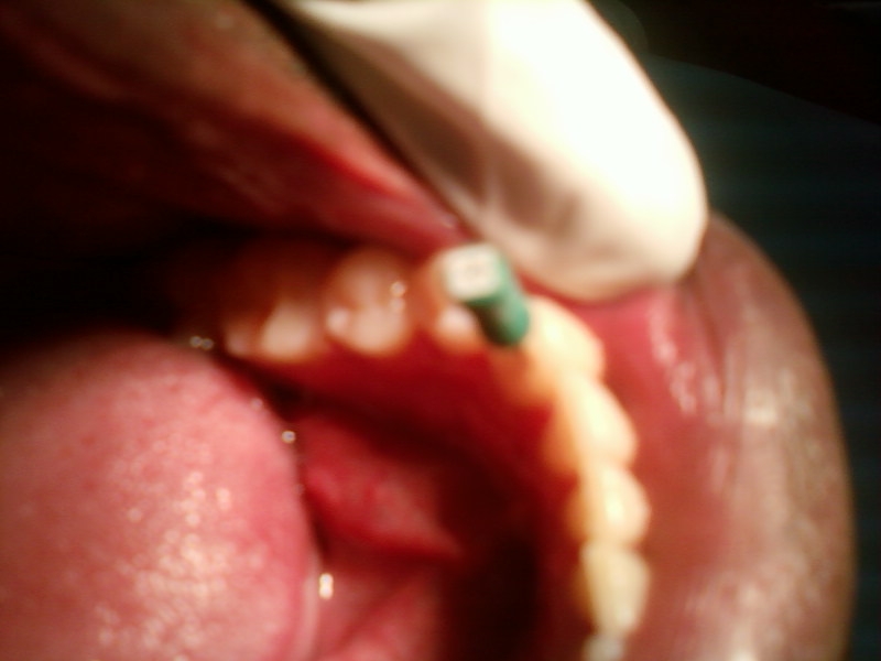
— Food debris are white small particles on the teeth- easily rinsed off
— Dental plaque is a thin film of bacteria that sticks to teeth – yellow in colour can’t be rinsed off, removed only by brushing & flossing
— Calculus is calcified plaque- hard, brownish or darkish in colour- only removed by scaling
Subscribe to:
Posts (Atom)